
How AI Medical Imaging Is Powering Precision Healthcare
Artificial intelligence is revolutionizing how medical images are acquired, analyzed, and interpreted. The transformation ushers in a new era of data-driven diagnostics and faster, more personalized patient care.
Artificial intelligence is revolutionizing how medical images are acquired, analyzed, and interpreted. The transformation ushers in a new era of data-driven diagnostics and faster, more personalized patient care.

Martin is a data scientist and AI engineer specializing in machine learning. After completing his Ph.D. in physics, he developed AI-powered tools and data products using diverse data types, including images, geospatial inputs, and natural language. He also built and deployed an end-to-end pipeline for medical imaging analysis, now in clinical use across Argentina, Brazil, and Chile.
Previous Role
Data ScientistPreviously At

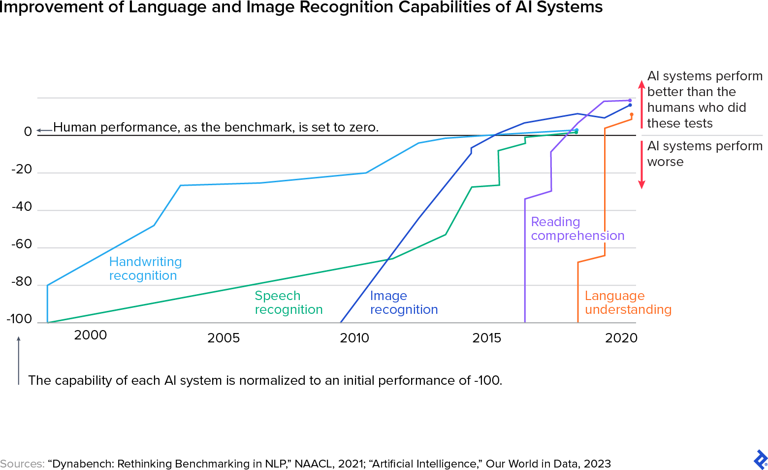
Radiology is widely regarded as the medical field most profoundly transformed by artificial intelligence. In fact, 76% of all FDA-approved AI algorithms to date focus on artificial intelligence in medical imaging, a clear sign of how central this technology has become. These advances are particularly prominent in computer vision, a subfield of AI focused on interpreting visual inputs, which has enabled radiology to evolve into a more precise, efficient, and scalable discipline. Computer vision now powers many core medical imaging tools, such as CT and MRI scans, X-rays, and ultrasounds.
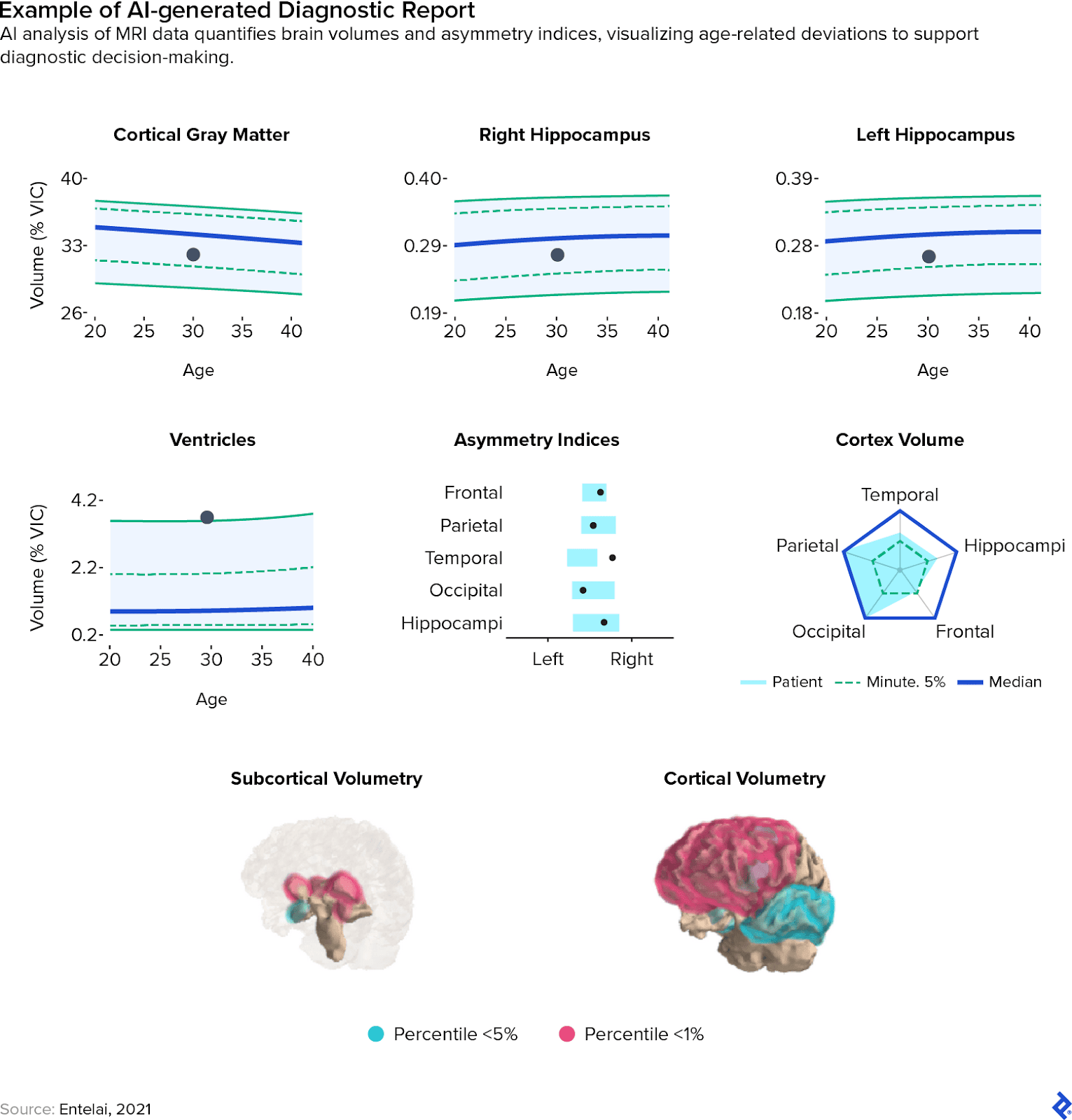
I’ve witnessed this transformation firsthand. As a lead data scientist at Entelai, an AI-and-healthcare startup, I helped develop and deploy a brain MRI analysis system that integrates directly into the imaging workflows at hospitals. This system automatically identifies and outlines demyelinating lesions—areas of nerve fiber damage commonly seen in conditions like multiple sclerosis—measures brain region volumes, and classifies patterns of atrophy, all while feeding results into patients’ electronic health records.
The journey of developing this system was not without its hurdles, but today, the system is in active use across multiple imaging centers in Latin America, with a steadily growing client base. This real-world experience, along with the technical and operational challenges we encountered, frames much of what this article explores: how AI in diagnostic imaging is reshaping the field, from acquisition to diagnosis.
Overview of AI in Medical Imaging
The rapid progress of medical AI imaging is due mainly to three key factors: the development of neural networks, the availability of large amounts of medical imaging data, and the power of graphics processing units (GPUs) for parallel computing.
At its core, neural networks are machine learning models made up of nodes (or “neurons”) that are interconnected in a layered arrangement. These networks learn to process information through a form of trial and error, adjusting their internal parameters based on feedback. Convolutional neural networks (CNNs), a subtype of neural networks, are especially well-suited for computer vision tasks due to their ability to extract spatial and hierarchical features from images. Each layer identifies a different aspect of the input—such as edges, shapes, or color gradients—building up a progressively detailed representation of the image.
Although CNNs were the focus of academic research for years, their breakthrough performance only became evident with the emergence of large, labeled image datasets. One landmark example is ImageNet, a dataset containing millions of annotated images organized according to a hierarchical structure. Datasets of this scale transformed computer vision, but training large CNNs on them introduced new computational challenges (including massive power demands). That’s where GPUs became essential. Designed for high-throughput parallel processing, GPUs dramatically reduced training times by handling the massive number of operations required to optimize neural networks. This made it possible to train and test models at a much faster pace.
Advancements like these have enabled technological feats that were unimaginable only a decade ago. Face tagging on social media, mobile check scanning, and image-based search have become routine parts of daily life. The same principles power AI and medical imaging applications, including X-rays, CT scans, and ultrasounds. AI imaging techniques are being developed to enhance MRI processes, including faster image reconstruction and advanced neuroimaging analysis, to improve efficiency and diagnostic capabilities. At Entelai, for example, we trained CNNs on curated, annotated brain MRI datasets to detect lesions and estimate brain region volumes. GPU acceleration was critical, particularly during the iterative process of improving segmentation accuracy.
AI medical imaging offers significant benefits: higher diagnostic accuracy, faster image processing, and lower overall costs. However, the sensitivity of healthcare environments means that automation must be approached with caution. Challenges and ethical considerations—such as transparency, bias mitigation, and clinical validation—must be integrated into every stage of product development.
Top 3 Benefits of Artificial Intelligence in Medical Imaging
Artificial intelligence in medical imaging helps radiologists work faster and more accurately, while reducing fatigue. This strengthens a workforce under significant strain, as the field currently faces a global talent shortage. Several factors contribute to this gap, including increased burnout among professionals and a surge in demand driven by an aging global population. Members of the scientific and medical communities are increasingly looking to imaging AI as a way to help alleviate this bottleneck by enabling radiologists to work more efficiently and with fewer fatigue-related errors. Below are three key ways AI medical imaging is moving the field toward a more scalable and resilient future.
1. AI Imaging Is a Quantitative Complement to Qualitative Medical Imaging Analysis
Radiologists rely on years of training and experience to interpret medical images and draw meaningful conclusions. The human eye excels at detecting nuanced patterns and integrating contextual information—such as patient medical history—into diagnostic reasoning. However, AI-powered computer vision systems are uniquely suited to extracting precise measurements and conducting large-scale quantitative analyses that would be time-prohibitive for human experts. For instance, delineating and calculating the volume of specific brain regions from an MRI scan is typically too labor-intensive to perform manually, but image analysis AI tools can complete this task rapidly and accurately.
In practice, at Entelai, we observed that AI-generated quantitative outputs helped radiologists validate their medical imaging analysis more confidently and consistently, even in fast-paced clinical environments. Rather than replacing expert judgment, AI imaging augments it with objective data and reproducible measurements.
2. AI Medical Image Analysis Reduces Error Rates and Improves Response Times
Fatigue from extended shifts increases the risk of diagnostic errors. AI medical imaging systems can act as reliable “second opinions” for radiologists, flagging potential oversights and prompting medical specialists to review ambiguous or high-risk findings. In addition, AI-powered triage tools can help optimize workflows by prioritizing urgent or complex cases for review early in a shift, when clinicians are most alert.
These systems can analyze variables such as shift schedules and historical performance data to assign cases more strategically. While approaches vary by implementation, triage-driven workflows have shown promising results in reducing diagnostic delays. For example, in a study focused on chest X-rays, AI prioritization reduced the average turnaround time for image reports from 11.2 days to just 2.7 days. Similar gains may be achievable across other imaging modalities as AI tools mature, translating to quicker diagnoses and earlier interventions for patients.
3. Reduced Costs and Round-the-Clock Availability
By automating repetitive tasks and streamlining image analysis, AI in diagnostic imaging has the potential to reduce operational costs. These efficiencies can make diagnostic services more affordable for healthcare systems and more accessible to patients. Unlike human radiologists, AI systems do not require rest and can provide uninterrupted support to care teams regardless of time or staffing constraints. This 24/7 capability is particularly valuable in remote or underserved regions, where access to expert interpretation is often limited.
3 Key Challenges of Using AI in Diagnostic Imaging
Despite its significant benefits, deploying AI for diagnostic purposes has ethical and technological challenges that must be carefully addressed. These are not abstract issues but rather ones with significant regulatory implications, especially as international frameworks such as GDPR and the EU AI Act increasingly set requirements for considerations like transparency and accountability. Below are three key hurdles facing AI in medical imaging today.
1. Dataset Bias
In curating a training dataset across clinics in Latin America, I saw firsthand how complex and critical this step can be. A machine learning model is only as reliable as the data it’s trained on. When datasets lack diversity or fail to reflect the populations they’re meant to serve, the resulting models can carry forward those biases. Research has demonstrated that this can lead to inaccurate diagnoses for underrepresented groups, exacerbating health disparities.
To avoid this, datasets should be built with deliberate attention to demographic and clinical diversity. Strategies like dataset rebalancing, data augmentation, and model recalibration can help mitigate bias, but these efforts must be part of a broader, ongoing commitment to ethical AI development. Our work at Entelai required a collaborative partnership with numerous clinics, engaging them in the creation of a comprehensive dataset, with the consent of individual patients who agreed to contribute to the data pool.
2. Model Generalization
For artificial intelligence in medical imaging to be clinically useful across different healthcare settings, it must reliably perform on new data from varied imaging equipment and patient populations. This ability, known as generalization, is one of the key challenges in developing diagnostic AI systems. In practical terms, generalization refers to a model’s capacity to apply what it has learned from one dataset to a separate, previously unseen set of data.
For example, a model trained on brain scans from one manufacturer’s MRI machines may not perform as well when applied to images from a different vendor, due to variations in resolution, contrast, or imaging protocols. These discrepancies can reduce diagnostic accuracy and undermine clinical trust. As a result, building robust, generalizable models requires not only diverse training data but also active validation across a range of sources.
We encountered this challenge during our deployment pipeline at Entelai. Variations in imaging equipment and acquisition settings across clinics occasionally led to drops in system performance. To manage this, we implemented a dedicated monitoring framework, vigilantly overseen by our quality assurance team, to track the integration of each new clinic. When significant performance degradation was detected, our team responded by fine-tuning existing models or developing dedicated ones calibrated to the specific data distribution of the new environment.
This experience underscored the importance of live monitoring, an essential component of responsible AI deployment. Monitoring helps detect underperformance early, allowing teams to intervene before inaccuracies reach patients or clinicians. In dynamic clinical settings, where real-world conditions often differ from training scenarios, adaptability and oversight are critical to maintaining model performance.
3. Lack of Explainability
AI medical imaging models often function as black boxes, meaning their decision-making processes can be opaque, even to the developers who built them. While these systems may deliver accurate results, their lack of interpretability presents a barrier to clinical trust and adoption. Clinicians and patients alike need to understand the rationale behind a diagnosis and not just accept it at face value.
To address this, researchers have developed a variety of explainability techniques, generally falling into three categories:
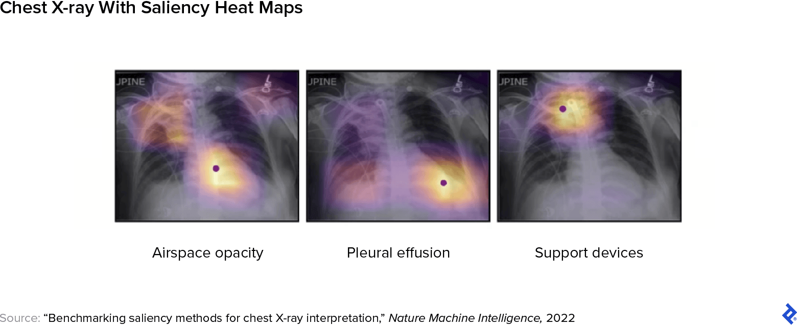
- Visual techniques, such as saliency maps, highlight the image regions that most influenced the model’s output, allowing clinicians to see where the model “looked” when forming its conclusion. For example, a saliency map superimposed on a CT scan can help pinpoint the area where the model detected an anomaly.
- Textual techniques generate concise explanations that describe the model’s reasoning in plain language. These are often implemented as decision support text, offering clinicians a written summary of the logic behind the output.
- Statistical techniques provide structured data—such as feature importance scores or model comparisons—that shed light on how predictions were made. Feature importance analysis identifies which image components most impacted the outcome, while model comparison allows teams to evaluate how different models perform on the same dataset.
Explainability is a rapidly evolving field, and improving transparency will be essential for building trust and ensuring safe, responsible deployment of AI in diagnostic workflows. The EU’s AI Act from 2024 includes an explicit regulatory requirement pertaining to transparency and interpretability in high-risk applications. By August 2026, these systems must be designed so that healthcare providers can interpret AI outputs and use them appropriately, supported by clear information on performance limitations and known risks.
How Accurate Is AI in Medical Imaging?
Research shows that in some applications, AI systems perform on par with—or even exceed—the accuracy of expert radiologists. In mammography screening, for example, a large population-based study found that AI matched the performance of double reading—a quality-control practice in which two independent radiologists review each scan—while also reducing workload by 44%. In a separate study on breast ultrasounds, AI systems reduced false positives by 37.3% and unnecessary biopsies by 27.8%.
Still, accuracy depends heavily on the use case and dataset quality. Researchers have found that models trained in controlled research environments can underperform when deployed in real-world clinical settings, where patient populations and image quality vary. This underscores the importance of human-AI collaboration in medical imaging: AI should be regarded as a complement to human expertise, pairing computational speed and pattern recognition with on-the-ground medical judgment. Ongoing performance validation and human oversight are essential to ensure reliable, context-appropriate results.
4 Steps to Modernize Radiological Workflows With AI Imaging
Every stage of the medical imaging workflow presents opportunities for improvement through AI. AI models are utilized to detect early signs of diseases such as breast cancer, lung cancer, tuberculosis, and cardiovascular risks. From the moment an image is acquired to the final reporting phase, AI technologies are helping radiologists work with greater efficiency and accuracy. Below is a step-by-step overview of how AI is being integrated into the radiological pipeline.
Step 1: Image Acquisition
The first step in any imaging workflow is acquiring a clean, high-quality image. Errors introduced here can cascade throughout the pipeline, leading to increased radiation exposure for patients or diagnostic delays. AI tools can now assist with minimizing these risks and improving acquisition efficiency in the most common medical imaging use cases.
- CT: Systems like Radiology Smart Assistant support automated patient positioning, aligning the body correctly within the scanner to ensure the region of interest is centered and within the field of view. These tools also perform error flagging, identifying issues such as motion artifacts in real time. This reduces unnecessary radiation exposure and improves scan efficiency.
- MRI: Siemens Healthineers applies deep learning to accelerate image acquisition and improve image reconstruction, a process that transforms raw signal data into clear, high-resolution images. AI can also automate slice positioning, deciding where in the body to acquire images and suggesting the appropriate scan protocol based on clinical context.
- Fluoroscopy: FluoroShield enables automated collimation, which dynamically adjusts the size and shape of the X-ray beam during procedures. This minimizes unnecessary radiation exposure to surrounding tissue and improves image focus on the area of interest, which is critical in dynamic studies of the gastrointestinal or cardiovascular systems.
- Ultrasound: AI-assisted ultrasound tools recognize anatomical landmarks in real time, guiding the sonographer to the correct imaging planes and automating organ identification. This is especially helpful in point-of-care settings, where scan quality can significantly affect diagnosis.
Step 2: Image Preprocessing
Once an image is captured, it must be transformed from raw data into a clinically usable format. This stage, known as preprocessing, involves noise reduction, artifact correction, and image reconstruction to enhance consistency and diagnostic quality. AI technologies have been especially impactful here, enabling faster and more accurate processing, even when scan conditions are less than ideal.
- CT: Tools like TrueFidelity use deep learning to reconstruct images from low-dose scans, preserving fine anatomical detail while minimizing radiation exposure. By learning from high-quality examples, these models can reduce artifacts and improve contrast, helping radiologists interpret subtle findings with greater confidence.
- MRI: AI is applied to accelerate MRI scan times and improve reconstruction fidelity, even with undersampled data. Deep learning models can recover missing spatial information and reduce noise, producing clearer images with sharper resolution, which is especially valuable for detecting small lesions or subtle structural changes.
Despite the positive implications for these use cases, it’s important to note that all image enhancement techniques carry some risk of introducing artifacts or over-smoothing regions, potentially masking abnormalities or generating details that were not present in the original scan. Robust validation and clinical oversight remain essential to ensure diagnostic reliability.
Step 3: Image Analysis and Interpretation
Once an image is preprocessed and ready for interpretation, the next step is analysis, which is arguably the phase where the combination of AI and medical imaging has the most exciting implications. Here, AI models assist with identifying abnormalities and classifying medical conditions, and can automate routine tasks like image segmentation, allowing radiologists to focus on more complex diagnostic challenges. These tasks form the basis for many clinical decisions and are typically the most labor-intensive for radiologists.
- Detection: This involves automatically identifying the presence and location of features such as tumors, fractures, or nodules within an image. AI medical image analysis models are trained to recognize specific patterns, like the density or shape of an abnormal mass, and can flag regions of interest with bounding boxes or markers. Detection tools can accelerate diagnosis time and reduce the risk of overlooking subtle or incidental findings.
- Segmentation: In this step, AI systems isolate anatomical structures or abnormalities by dividing an image into labeled regions. For example, segmentation models can outline brain ventricles, delineate tumor margins, or separate lung lobes, often down to the pixel level. This enables precise measurements, volumetric analysis, and 3D reconstructions, which are critical for treatment planning, surgical navigation, and disease monitoring.
- Classification: After detection and segmentation, AI can assign diagnostic labels to findings, such as determining whether a lesion is benign or malignant, or classifying tissue changes consistent with disease progression. Classification models often incorporate probabilistic scoring, giving clinicians a confidence level that can guide further investigation or action.
The detection, segmentation, and classification functions underpin a wide range of clinical applications for artificial intelligence in medical imaging:
- X-ray: AI tools for chest X-rays can detect conditions like pneumonia, pleural effusion, or cardiomegaly, often with accuracy comparable to human radiologists. In mammography, AI is being deployed as a second reader to identify subtle signs of breast cancer and reduce false negatives. These systems help triage high-risk cases and provide decision support in settings with limited radiology expertise.
- CT: During the COVID-19 pandemic, AI-driven CT analysis emerged as a powerful tool for detecting pulmonary hallmarks of the disease, enabling clinicians to triage patients in high-pace, high-volume clinical settings. These tools also supported monitoring of disease severity and assessment of long-term lung changes, which often persist months after recovery. Separately, in dental imaging, fully automatic AI systems have been integrated with cone-beam CT (CBCT) to delineate dental structures, achieving segmentation accuracy comparable to that of experienced radiologists, while operating up to 500 times faster.
- MRI: In neuroimaging, AI tools like FastSurfer use deep neural networks to accelerate brain segmentation workflows by automating the identification of regions affected by conditions such as multiple sclerosis. Meanwhile, the AI system we developed at Entelai enabled automated segmentation of cortical and subcortical brain regions, supporting clinical diagnosis and the longitudinal tracking of neurodegeneration.
- Ultrasound: AI technologies are being used to improve consistency and diagnostic accuracy in a variety of ultrasound applications, including breast imaging and echocardiography. In particular, deep learning can guide image acquisition and automate interpretation, reducing reliance on operator expertise and expanding access to high-quality care, even in resource-limited or remote settings.
Step 4: Reporting and Clinical Communication
The final step in the radiological workflow is the generation of structured reports, which synthesize findings into actionable insights for referring physicians. These reports are highly specialized, often dense with clinical terminology, and must balance precision with efficiency, especially in high-volume settings. Traditionally, radiologists dictate or manually enter findings, but this process can be time-consuming and prone to inconsistencies.
AI is beginning to transform this phase by supporting both report generation and comprehension. Natural language processing (NLP) tools can automatically extract key information from image analyses and draft preliminary reports, reducing documentation burden and standardizing terminology. While not infallible, these systems can support consistency and speed when used alongside expert review. Advanced systems are also exploring report simplification, translating technical findings into accessible language for nonspecialist physicians or patients. As these capabilities evolve, they hold the potential to streamline communication and improve continuity of care across clinical teams.
Understanding the Building Blocks of AI in Diagnostic Imaging
AI models vary based on the type of input they process—such as images, text, audio, or sensor data—and the kind of output they’re designed to produce. Many clinical tools combine multiple models into a pipeline, or even into multimodal systems that integrate vision, language, and real-time sensor feedback.
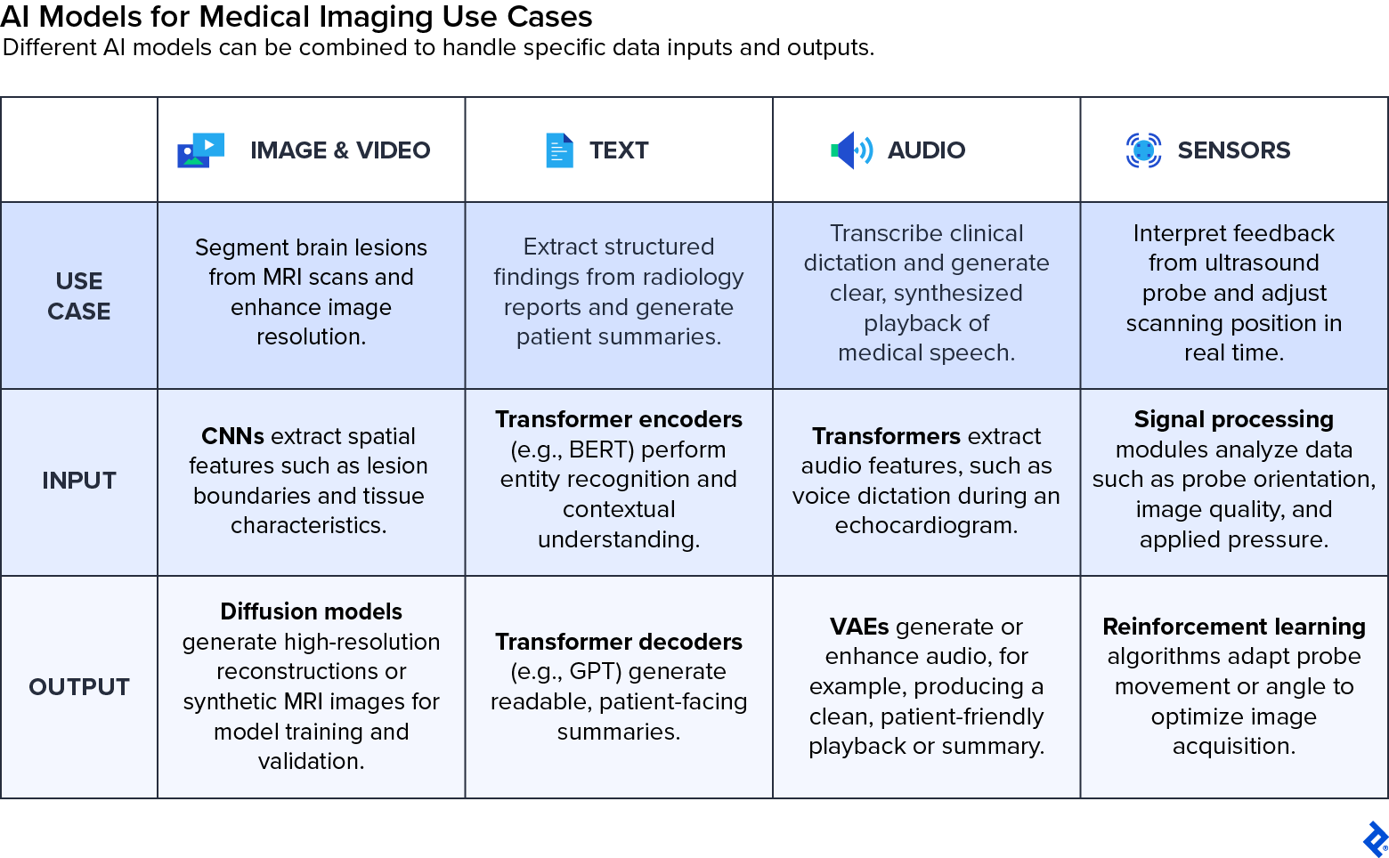
Below is an overview of the main types of medical imaging AI models and how they’re used:
- Image and video models are at the core of radiology AI, including the system we developed at Entelai. CNNs are commonly used for classification and segmentation tasks, such as detecting lung nodules or measuring organ volumes. More advanced architectures like vision transformers capture broader spatial context, while generative models, such as generative adversarial networks (GANs), diffusion networks, and variational autoencoders (VAEs), are increasingly used for image enhancement, synthetic data generation, and style transfer.
- Text models help structure and interpret the written components of radiology workflows. Transformer-based models like GPT and BERT (short for bidirectional encoder representations from transformers) can summarize reports, extract structured findings, or even translate technical imaging descriptions into patient-friendly language. Other models use word embeddings to capture the meaning of medical terms based on context, or long short-term memory networks (LSTMs) to process text sequences such as radiology notes over time. It’s important to note that text models can occasionally generate hallucinations, so healthcare providers must always review their outputs carefully to ensure clinical accuracy.
- Audio models are applied in clinical environments for signal processing, speech recognition, and voice command functionality. These include recurrent neural networks (RNNs), LSTMs, and transformers that power real-time transcription tools, enabling radiologists to dictate findings hands-free. In addition to transcription, traditional speech synthesis techniques and variational autoencoders are used to create or enhance clinical audio, such as generating natural-sounding speech or filtering out noise from recordings.
- Sensor and actuator models interpret real-time input from physical devices, such as ultrasound probes or robotic instruments, and generate output signals that guide motion or positioning. These models rely on signal processing, control theory, and reinforcement learning to continuously adjust actions based on sensor feedback, improving precision and reducing the need for manual correction during procedures.
As medical imaging AI systems grow more sophisticated, these model types are increasingly integrated across modalities. For example, a multimodal platform might combine brain MRI segmentation, clinical text analysis, and longitudinal tracking to support more holistic assessments of neurodegenerative diseases. This convergence is opening the door to diagnostic systems that synthesize diverse clinical data streams to deliver broader patient-centered insight.
What Are the Future Trends of AI in Medical Imaging?
The current wave of medical imaging AI has been driven largely by advances in computer vision, with additional gains from natural language processing for report generation and structured documentation. But a new chapter is beginning—one focused on multimodal AI systems that can integrate a variety of inputs, including medical images, clinical text, sensor data, and patient history, into a unified diagnostic model. The developments point toward the advent of generalist medical AI models capable of transforming the entire diagnostic process.
These next-generation systems aim to move beyond analyzing a single scan in isolation. Instead, they can synthesize information across modalities and time, factoring in prior imaging, lab results, and physician notes to generate more comprehensive and clinically useful reports. This shift mirrors how human clinicians reason, drawing from multiple data points to inform judgment rather than relying solely on one image or test.
During my time at Entelai, we took foundational steps in this direction. We integrated automated brain MRI analysis with electronic health records to help radiologists interpret new findings in the context of quantitative trends over time. As these capabilities evolve, they offer a glimpse of how AI could move beyond image interpretation to begin supporting broader clinical reasoning.
To realize this potential, technical progress must be matched by careful validation and ethical deployment. The deployment of AI in medical imaging is influenced by regulatory hurdles and the need for transparent algorithms that build trust and ensure accountability. Issues like explainability, regulatory approval, dataset bias, and the need for high-quality, labeled data to ensure accurate and equitable outcomes will shape how quickly and responsibly these systems reach widespread adoption. Educational initiatives and summits are exploring these ethical considerations as well as the legal aspects and training requirements for integrating AI into radiology practices.
Integrations must emphasize the importance of human expertise, where artificial intelligence in medical imaging aids but does not replace radiologists, enhancing diagnostic accuracy and efficiency while preserving high-quality patient care and human clinical judgment. If done right, this next phase of AI imaging could transform diagnostics from a static snapshot into a continuously learning, decision-support ecosystem that delivers richer and more personalized care at scale.
The technical content presented in this article was reviewed by Roman Vlasov.
Further Reading on the Toptal Blog:
Understanding the basics
The use of AI in medical imaging enables greater accuracy and efficiency while reducing diagnostic turnaround time. It powers key technologies, such as CT scans, MRIs, X-rays, and ultrasounds, and provides enhanced image quality and automated interpretation. AI also supports early detection through pattern recognition and predictive analytics.
AI was first explored in medical imaging in the 1960s, primarily for tasks like pattern recognition. One early example was the Film Input to Digital Automatic Computer (FIDAC), developed to digitize and interpret medical images. The field advanced significantly in the 2010s with the rise of deep learning and access to large annotated datasets, enabling more accurate and scalable diagnostic tools.
AI is used in CT scans to optimize image acquisition and enhance diagnostic quality. It reduces noise and artifacts through advanced reconstruction techniques and supports detailed segmentation of tissues and organs, helping to flag abnormalities. By accelerating diagnosis through automated medical image analysis, AI minimizes the need for rescans and therefore reduces radiation exposure for patients.
While AI is transforming radiology by improving diagnostic accuracy and accelerating image interpretation, it is not expected to replace radiologists. Instead, AI is increasingly being used as a decision-support tool that enhances human expertise in patient care rather than replacing it.
Buenos Aires, Argentina
Member since December 11, 2020
About the author
Martin is a data scientist and AI engineer specializing in machine learning. After completing his Ph.D. in physics, he developed AI-powered tools and data products using diverse data types, including images, geospatial inputs, and natural language. He also built and deployed an end-to-end pipeline for medical imaging analysis, now in clinical use across Argentina, Brazil, and Chile.
Previous Role
Data ScientistPREVIOUSLY AT